
What type of preclinical studies do you require?


Drug development

Tissue regeneration, new biomaterials, stem cells

Medical devices
-
0
preclinical trials
-
0
therapeutic areas
-
0
models
Therapeutic areas of expertise
 / 12
/ 12
Dermatology
PK, TK, PD, TOX // Device implantation // Wound healing // Impaired wound healing // Burn model // Wounds with and without challenge // Different wound depth and dimensions // MRI imaging
Robotic surgery
Robot validation // Usability tests // Laparoscopy // Ophthalmology // Neurosurgery
 / 12
/ 12
Laparoscopic
PK, TK, PD, TOX // Device implantation // Laparoscopic administration // Manual and robotic surgery
 / 12
/ 12
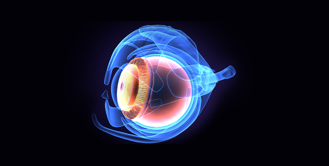
Ophthalmology
PK, TK, PD, TOX // Macular degeneration model // Intravitreal, subretinal and suprachoroidal injections // Vitrectomy // ERG, IOP, OCT
Digestive system
PK, TK, PD, TOX // Device implantation // Endoscopic and laparoscopic administration and surgery
 / 12
/ 12
Dental
Biomaterials // Bone regeneration // Dental implants // Maxillofacial surgery // CT Scan imaging
 / 12
/ 12
Cardiovascular
PK, TK, PD, TOX // Device implantation // Thrombus model // Myocardial Infarct model
 / 12
/ 12
Neonatal
PK, TK, PD, TOX // Preterm and neonatal // NEC model
 / 12
/ 12
Renal
Device implantation // Open and laparoscopic surgery // Renal transplant // Partial and total nephrectomy
Respiratory system
Device Implantation // ICU care model
 / 12
/ 12
Neurosurgery
PK, TK, PD, TOX // Device implantation // Intracranial administration // MRI imaging
 / 12
/ 12
Sepsis
PK, TK, PD, TOX // Sepsis model
Preclinical models
Preclinical animal models
Miniature pigs: Specipig, Göttingen minipig.
Other preclinical models
Efficient models for innovative outcomes
If something can be imagined, it can be created, just like our ad hoc open models.
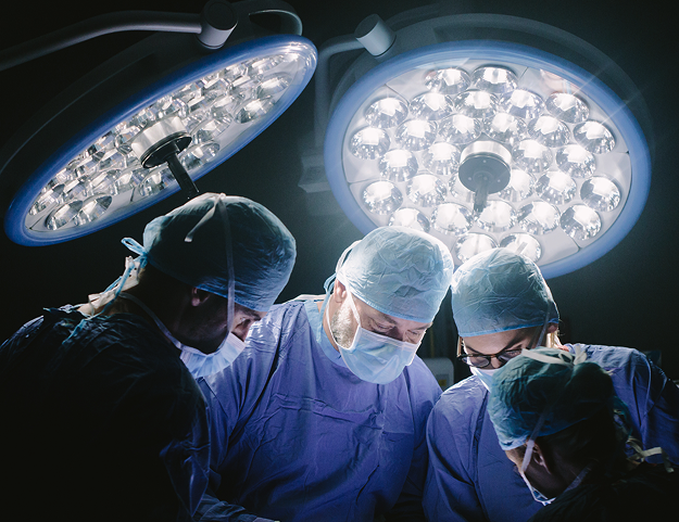
With our network of experts and surgeons, we can create a wide variety of surgical models across different systems and organs. Furthermore, we are equipped to perform the most advanced surgical techniques and procedures. Some of the disease preclinical models developed so far at Versa Biomedical include:


Skin-Dermatology
Wound healing model / Wound challenge model / Skin inflammation model / Skin burn model / Full and partial wound healing model / SC administration

Eyes – Ophthalmology
Macular degeneration model / Blindness induction model / Behavioural eye vision labyrinth

Renal
Renal transplant model / Partial or total nephrectomy model / Cold and warm ischemia model

Sepsis
LPS sepsis model / Acute clinical sepsis model / Acute subclinical sepsis model

Neonate – Preterm
Preterm UCI necrotic enterocolitis (NEC) model

Dental
Implant model / Dental bone regeneration

Brain
Ischemic stroke model

Cardiovascular
Thrombus model / Myocardial infarct (MI) / ischemia model

OncoSpecipig
We are developing a Specipig hybrid GM oncology model together with Sus Clinicals

I+D
Research and Development of new disease pig models / Customization of new disease or surgical pig models
Experts in designing flexible, biomedical models


Based on our expertise and in collaboration with leading experts in each therapeutic area, we can develop a proposal for the pig model that best aligns with the preclinical research aims. To achieve these, we work based on experience, bibliography or information provided by the sponsor or client.
Related projects
Stay up to date with all Versa Biomedical news, events and achievements.
A swine model of selective geographic atrophy of outer retinal layers mimicking atrophic AMD: A phase I escalating dose of subretinal sodium iodate
Despite recent advances in treatment of the wet form of AMD,1–6 the clinical outcome of AMD often remains blindness. Nowadays, the great challenge to which the ophthalmologic community is committed lies in finding a treatment that may slow the relentless progression of the atrophic form of the disease, together with other approaches that restore or regenerate the involved part of the diseased retinal tissue. Nowadays, the advanced form of dry AMD with geographic atrophy (GA) may be the first cause of legal blindness among elderly patients in the industrialized world, and it represents up to a third of cases of late AMD.7,8 Besides the impact of the disease among individuals, the economic burden of atrophic AMD (drusen, RPE abnormalities, and/or GA) in the United States is enormous: $26,100 million annually in one study9 and approximately 0.22% of its gross domestic product in 2010 in terms of wage loss from untreated disease in another report.10
Download case
Abstract of the publication:
Purpose
To establish the dose of subretinal sodium iodate (NaIO3) in order to create a toxin-induced large animal model of selective circumscribed atrophy of outer retinal layers, the retinal pigment epithelium (RPE), and photoreceptors, by spectral-domain optical coherence tomography (SD-OCT) and immunocytochemistry.Methods
Fifteen male and female healthy Yorkshire pigs received unilateral subretinal escalating doses of NaIO3 under general anesthesia. In all the animals, volumes of 0.1 to 0.2 mL NaIO3 were injected into the subretinal space of the area centralis through a 23/38-gauge subretinal cannula. Control SD-OCTs were performed 1 and 2 months after the surgery, at which time pigs were euthanized and eyes enucleated. Globes were routinely processed for histologic and immunohistochemical evaluation.Results
Spectral-domain OCT and immunohistochemistry revealed circumscribed and well-demarcated funduscopic lesions, limited to the outer retinal layers in pigs treated with 0.01 mg/mL subretinal sodium iodate.Conclusions
The swine model of a controlled area of circumscribed retinal damage, with well-delimited borders, and selectively of the outer layers of the retina presented herein shows several clinical and histologic features of geographic atrophy in AMD. Therefore, it may represent a valuable tool in the investigation of new emerging regenerative therapies that aim to restore visual function, such as stem cell transplantation or optogenetics.
Dental implant research
Despite recent advances in treatment of the wet form of AMD,1–6 the clinical outcome of AMD often remains blindness. Nowadays, the great challenge to which the ophthalmologic community is committed lies in finding a treatment that may slow the relentless progression of the atrophic form of the disease, together with other approaches that restore or regenerate the involved part of the diseased retinal tissue. Nowadays, the advanced form of dry AMD with geographic atrophy (GA) may be the first cause of legal blindness among elderly patients in the industrialized world, and it represents up to a third of cases of late AMD.7,8 Besides the impact of the disease among individuals, the economic burden of atrophic AMD (drusen, RPE abnormalities, and/or GA) in the United States is enormous: $26,100 million annually in one study9 and approximately 0.22% of its gross domestic product in 2010 in terms of wage loss from untreated disease in another report.10
Download case
Abstract of the publication:
Purpose
To establish the dose of subretinal sodium iodate (NaIO3) in order to create a toxin-induced large animal model of selective circumscribed atrophy of outer retinal layers, the retinal pigment epithelium (RPE), and photoreceptors, by spectral-domain optical coherence tomography (SD-OCT) and immunocytochemistry.
Methods
Fifteen male and female healthy Yorkshire pigs received unilateral subretinal escalating doses of NaIO3 under general anesthesia. In all the animals, volumes of 0.1 to 0.2 mL NaIO3 were injected into the subretinal space of the area centralis through a 23/38-gauge subretinal cannula. Control SD-OCTs were performed 1 and 2 months after the surgery, at which time pigs were euthanized and eyes enucleated. Globes were routinely processed for histologic and immunohistochemical evaluation.
Results
Spectral-domain OCT and immunohistochemistry revealed circumscribed and well-demarcated funduscopic lesions, limited to the outer retinal layers in pigs treated with 0.01 mg/mL subretinal sodium iodate.
Conclusions
The swine model of a controlled area of circumscribed retinal damage, with well-delimited borders, and selectively of the outer layers of the retina presented herein shows several clinical and histologic features of geographic atrophy in AMD. Therefore, it may represent a valuable tool in the investigation of new emerging regenerative therapies that aim to restore visual function, such as stem cell transplantation or optogenetics.

Establishment of a reproducible and minimally invasive ischemic stroke model in swine
Despite recent advances in treatment of the wet form of AMD,1–6 the clinical outcome of AMD often remains blindness. Nowadays, the great challenge to which the ophthalmologic community is committed lies in finding a treatment that may slow the relentless progression of the atrophic form of the disease, together with other approaches that restore or regenerate the involved part of the diseased retinal tissue. Nowadays, the advanced form of dry AMD with geographic atrophy (GA) may be the first cause of legal blindness among elderly patients in the industrialized world, and it represents up to a third of cases of late AMD.7,8 Besides the impact of the disease among individuals, the economic burden of atrophic AMD (drusen, RPE abnormalities, and/or GA) in the United States is enormous: $26,100 million annually in one study9 and approximately 0.22% of its gross domestic product in 2010 in terms of wage loss from untreated disease in another report.10
Download case

Abstract of the publication:
Purpose
To establish the dose of subretinal sodium iodate (NaIO3) in order to create a toxin-induced large animal model of selective circumscribed atrophy of outer retinal layers, the retinal pigment epithelium (RPE), and photoreceptors, by spectral-domain optical coherence tomography (SD-OCT) and immunocytochemistry.Methods
Fifteen male and female healthy Yorkshire pigs received unilateral subretinal escalating doses of NaIO3 under general anesthesia. In all the animals, volumes of 0.1 to 0.2 mL NaIO3 were injected into the subretinal space of the area centralis through a 23/38-gauge subretinal cannula. Control SD-OCTs were performed 1 and 2 months after the surgery, at which time pigs were euthanized and eyes enucleated. Globes were routinely processed for histologic and immunohistochemical evaluation.Results
Spectral-domain OCT and immunohistochemistry revealed circumscribed and well-demarcated funduscopic lesions, limited to the outer retinal layers in pigs treated with 0.01 mg/mL subretinal sodium iodate.Conclusions
The swine model of a controlled area of circumscribed retinal damage, with well-delimited borders, and selectively of the outer layers of the retina presented herein shows several clinical and histologic features of geographic atrophy in AMD. Therefore, it may represent a valuable tool in the investigation of new emerging regenerative therapies that aim to restore visual function, such as stem cell transplantation or optogenetics.
Specipig miniature pig in metabolic disease insulin independent SC administration PK study
Despite recent advances in treatment of the wet form of AMD,1–6 the clinical outcome of AMD often remains blindness. Nowadays, the great challenge to which the ophthalmologic community is committed lies in finding a treatment that may slow the relentless progression of the atrophic form of the disease, together with other approaches that restore or regenerate the involved part of the diseased retinal tissue. Nowadays, the advanced form of dry AMD with geographic atrophy (GA) may be the first cause of legal blindness among elderly patients in the industrialized world, and it represents up to a third of cases of late AMD.7,8 Besides the impact of the disease among individuals, the economic burden of atrophic AMD (drusen, RPE abnormalities, and/or GA) in the United States is enormous: $26,100 million annually in one study9 and approximately 0.22% of its gross domestic product in 2010 in terms of wage loss from untreated disease in another report.10
Download case
