
Each client is unique. So is the team needed to carry out their project


Thanks to our extensive network and versatility, we provide the best experts in every field, both in human or veterinary medicine. We also welcome surgeons and experts provided by sponsors to assist with the projects.
Our approach involves an ad hoc team, a tailored model and solid results. We are open and flexible because we value the uniqueness of each case and we thrive on accessible innovation.
Ready to achieve your project goals
Such an extensive network of experts in different therapeutic areas offers multiple advantages. Amongst them:
 Widen our expertise areas
 Extend our capabilities
 Reduce risks
 Ensure data relevance
 Accelerate timings
 Leverage experience portfolio
The role of experts in our work process
Our network of experts and collaborating partners, with their expertise, complements and enriches the work of our team to achieve our sponsors' objectives in the most efficient way possible. Including both the protocol preparation and the study execution.
After defining the protocol to follow and gathering the necessary authorizations to carry out a specific project, we enter the phase of inclusion and execution of the study.
-
01
Protocol & AuthorizationAnimal Welfare Body & Ethical Comitte
-
02
Inclusion + Study execution
Surgeon and other experts
Histopathology, Bioanalysis, Specific test or imaging
-
03
Final reportDelegated phase final reports
Meet our collaborators
We increasingly have more specialized professionals when assembling the ad hoc teams that carry out each project we develop. These are the professionals and partners we have collaborated with so far:
Please specify the types of professionals or therapeutic areas that interest you:


Fuco Novoa

Dr Santiago Candela Canto, PhD

Dr Josep Rubio Palau

Dra Marta Leiva Repiso


Txema Castelar Cifuentes

Dr García Linares


Dr Roland Glauser
Magnus Persson

Dr Lluís Peri
Mª Teresa Peña Giménez
Peter-Paul Fransen

Geert van Almen

Montse Jorro

Teresa Gasull
Juan M Carretero Bellón

Prof Jordi Monés
02 / Partnerships
Pioneering companies in the field of health:


Related projects
Stay up to date with all Versa Biomedical news, events and achievements.
A swine model of selective geographic atrophy of outer retinal layers mimicking atrophic AMD: A phase I escalating dose of subretinal sodium iodate
Despite recent advances in treatment of the wet form of AMD,1–6 the clinical outcome of AMD often remains blindness. Nowadays, the great challenge to which the ophthalmologic community is committed lies in finding a treatment that may slow the relentless progression of the atrophic form of the disease, together with other approaches that restore or regenerate the involved part of the diseased retinal tissue. Nowadays, the advanced form of dry AMD with geographic atrophy (GA) may be the first cause of legal blindness among elderly patients in the industrialized world, and it represents up to a third of cases of late AMD.7,8 Besides the impact of the disease among individuals, the economic burden of atrophic AMD (drusen, RPE abnormalities, and/or GA) in the United States is enormous: $26,100 million annually in one study9 and approximately 0.22% of its gross domestic product in 2010 in terms of wage loss from untreated disease in another report.10
Download case
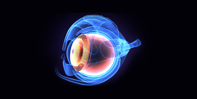
Abstract of the publication:
Purpose
To establish the dose of subretinal sodium iodate (NaIO3) in order to create a toxin-induced large animal model of selective circumscribed atrophy of outer retinal layers, the retinal pigment epithelium (RPE), and photoreceptors, by spectral-domain optical coherence tomography (SD-OCT) and immunocytochemistry.Methods
Fifteen male and female healthy Yorkshire pigs received unilateral subretinal escalating doses of NaIO3 under general anesthesia. In all the animals, volumes of 0.1 to 0.2 mL NaIO3 were injected into the subretinal space of the area centralis through a 23/38-gauge subretinal cannula. Control SD-OCTs were performed 1 and 2 months after the surgery, at which time pigs were euthanized and eyes enucleated. Globes were routinely processed for histologic and immunohistochemical evaluation.Results
Spectral-domain OCT and immunohistochemistry revealed circumscribed and well-demarcated funduscopic lesions, limited to the outer retinal layers in pigs treated with 0.01 mg/mL subretinal sodium iodate.Conclusions
The swine model of a controlled area of circumscribed retinal damage, with well-delimited borders, and selectively of the outer layers of the retina presented herein shows several clinical and histologic features of geographic atrophy in AMD. Therefore, it may represent a valuable tool in the investigation of new emerging regenerative therapies that aim to restore visual function, such as stem cell transplantation or optogenetics.
Dental implant research
Despite recent advances in treatment of the wet form of AMD,1–6 the clinical outcome of AMD often remains blindness. Nowadays, the great challenge to which the ophthalmologic community is committed lies in finding a treatment that may slow the relentless progression of the atrophic form of the disease, together with other approaches that restore or regenerate the involved part of the diseased retinal tissue. Nowadays, the advanced form of dry AMD with geographic atrophy (GA) may be the first cause of legal blindness among elderly patients in the industrialized world, and it represents up to a third of cases of late AMD.7,8 Besides the impact of the disease among individuals, the economic burden of atrophic AMD (drusen, RPE abnormalities, and/or GA) in the United States is enormous: $26,100 million annually in one study9 and approximately 0.22% of its gross domestic product in 2010 in terms of wage loss from untreated disease in another report.10
Download case
Abstract of the publication:
Purpose
To establish the dose of subretinal sodium iodate (NaIO3) in order to create a toxin-induced large animal model of selective circumscribed atrophy of outer retinal layers, the retinal pigment epithelium (RPE), and photoreceptors, by spectral-domain optical coherence tomography (SD-OCT) and immunocytochemistry.
Methods
Fifteen male and female healthy Yorkshire pigs received unilateral subretinal escalating doses of NaIO3 under general anesthesia. In all the animals, volumes of 0.1 to 0.2 mL NaIO3 were injected into the subretinal space of the area centralis through a 23/38-gauge subretinal cannula. Control SD-OCTs were performed 1 and 2 months after the surgery, at which time pigs were euthanized and eyes enucleated. Globes were routinely processed for histologic and immunohistochemical evaluation.
Results
Spectral-domain OCT and immunohistochemistry revealed circumscribed and well-demarcated funduscopic lesions, limited to the outer retinal layers in pigs treated with 0.01 mg/mL subretinal sodium iodate.
Conclusions
The swine model of a controlled area of circumscribed retinal damage, with well-delimited borders, and selectively of the outer layers of the retina presented herein shows several clinical and histologic features of geographic atrophy in AMD. Therefore, it may represent a valuable tool in the investigation of new emerging regenerative therapies that aim to restore visual function, such as stem cell transplantation or optogenetics.

Establishment of a reproducible and minimally invasive ischemic stroke model in swine
Despite recent advances in treatment of the wet form of AMD,1–6 the clinical outcome of AMD often remains blindness. Nowadays, the great challenge to which the ophthalmologic community is committed lies in finding a treatment that may slow the relentless progression of the atrophic form of the disease, together with other approaches that restore or regenerate the involved part of the diseased retinal tissue. Nowadays, the advanced form of dry AMD with geographic atrophy (GA) may be the first cause of legal blindness among elderly patients in the industrialized world, and it represents up to a third of cases of late AMD.7,8 Besides the impact of the disease among individuals, the economic burden of atrophic AMD (drusen, RPE abnormalities, and/or GA) in the United States is enormous: $26,100 million annually in one study9 and approximately 0.22% of its gross domestic product in 2010 in terms of wage loss from untreated disease in another report.10
Download case
Abstract of the publication:
Purpose
To establish the dose of subretinal sodium iodate (NaIO3) in order to create a toxin-induced large animal model of selective circumscribed atrophy of outer retinal layers, the retinal pigment epithelium (RPE), and photoreceptors, by spectral-domain optical coherence tomography (SD-OCT) and immunocytochemistry.Methods
Fifteen male and female healthy Yorkshire pigs received unilateral subretinal escalating doses of NaIO3 under general anesthesia. In all the animals, volumes of 0.1 to 0.2 mL NaIO3 were injected into the subretinal space of the area centralis through a 23/38-gauge subretinal cannula. Control SD-OCTs were performed 1 and 2 months after the surgery, at which time pigs were euthanized and eyes enucleated. Globes were routinely processed for histologic and immunohistochemical evaluation.Results
Spectral-domain OCT and immunohistochemistry revealed circumscribed and well-demarcated funduscopic lesions, limited to the outer retinal layers in pigs treated with 0.01 mg/mL subretinal sodium iodate.Conclusions
The swine model of a controlled area of circumscribed retinal damage, with well-delimited borders, and selectively of the outer layers of the retina presented herein shows several clinical and histologic features of geographic atrophy in AMD. Therefore, it may represent a valuable tool in the investigation of new emerging regenerative therapies that aim to restore visual function, such as stem cell transplantation or optogenetics.
Specipig miniature pig in metabolic disease insulin independent SC administration PK study
Despite recent advances in treatment of the wet form of AMD,1–6 the clinical outcome of AMD often remains blindness. Nowadays, the great challenge to which the ophthalmologic community is committed lies in finding a treatment that may slow the relentless progression of the atrophic form of the disease, together with other approaches that restore or regenerate the involved part of the diseased retinal tissue. Nowadays, the advanced form of dry AMD with geographic atrophy (GA) may be the first cause of legal blindness among elderly patients in the industrialized world, and it represents up to a third of cases of late AMD.7,8 Besides the impact of the disease among individuals, the economic burden of atrophic AMD (drusen, RPE abnormalities, and/or GA) in the United States is enormous: $26,100 million annually in one study9 and approximately 0.22% of its gross domestic product in 2010 in terms of wage loss from untreated disease in another report.10
Download case
