

Specipig miniature pig in metabolic disease insulin independent SC administration PK study
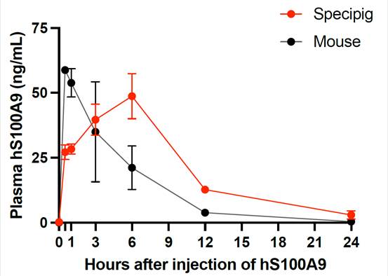
A pharmacokinetic (PK) assessment was performed in a porcine model (Specipig miniature pig), which was expected to greatly predict PK behaviour of a recombinant S100A9 in the reduction of therapeutic insulin needs after subcutaneous injection in humans. This model showed much greater plasma bioavailability of S100A9 compared to the one obtained at the same dose in the mouse model.
 Download case
Abstract of the publication:
Type 1 diabetes mellitus (T1DM) is characterized by insulin deficiency leading to hyperglycemia and several metabolic defects. Insulin therapy remains the cornerstone of T1DM management, yet it increases the risk of life-threatening hypoglycemia and the development of major comorbidities. Here, we report an insulin signaling–independent pathway able to improve glycemic control in T1DM rodents. Co-treatment with recombinant S100 calcium-binding protein A9 (S100A9) enabled increased adherence to glycemic targets with half as much insulin and without caus- ing hypoglycemia. Mechanistically, we demonstrate that the hyperglycemia-suppressing action of S100A9 is due to a Toll-like receptor 4–dependent increase in glucose uptake in specific skeletal muscles (i.e., soleus and dia- phragm). In addition, we found that T1DM mice have abnormal systemic inflammation, which is resolved by S100A9 therapy alone (or in combination with low insulin), hence uncovering a potent anti-inflammatory action of S100A9 in T1DM. In summary, our findings reveal the S100A9-TLR4 skeletal muscle axis as a promising therapeutic target for improving T1DM treatment.
Establishment of a reproducible and minimally invasive ischemic stroke model in swine
Researchers of the Cellular and Molecular Neurobiology (CMN) Research Group at the Germans Trias i Pujol Institute (IGTP) have developed and established a novel, reproducible and minimally invasive stroke model in the pig through an endovascular approach. The work has been conducted at the Centre for Comparative Medicine and Bioimage (CMCiB), a centre devoted to translational medicine at the very core of the Can Ruti Campus in Badalona together with the IGTP and the Germans Trias i Pujol University Hospital.

“Being able to reproduce the damage in specific brain areas in the model in Specipig’s pig breed and conventional pigs is important to any study that aims to determine the true neuroprotective effect of new molecules to be tested in brains similar to those of humans.”
Teresa Gasull, Senior Research Investigator & Team leader of the CMN group
Abstract of the publication:
The need for advances in the management/treatment options for ischemic stroke patients requires that upcoming preclinical research uses animals with more human-like brain characteristics. The porcine brain is considered appropriate, although the presence of the rete mirabile (RM) prevents direct catheterization of the intracranial arteries to produce focal cerebral ischemia. To develop a reproducible minimally invasive porcine stroke model, a guide catheter and guide wire were introduced through the femoral artery until reaching the left RM. Using the pressure cooker technique, Squid-12 embolization material was deposited to fill, overflow, and occlude the left RM, the left internal carotid artery, and left circle of Willis wing up to the origins of the middle cerebral arteries (MCAs), mimicking the occlusion produced in the filament model in rodents. Longitudinal multimodal cerebral MRI was conducted to assess the brain damage and cerebral blood supply. The technique we describe here occluded up to the origins of the MCAs in 7 of 8 swine, inducing early damage 90 minutes after occlusion that later evolved to a large cerebral infarction and producing no mortality during the intervention. This minimally invasive ischemic stroke model in swine produced reproducible infarcts and shows translational features common to human stroke.Keywords:
Neuroscience; Stroke.
Dental implant research
Specipig has signed an agreement with Zircon Medical, Swiss-based ceramic implant manufacturer made up of a team of seasoned experts in dentistry who are passionate about improving the quality of life of patients worldwide by making Patent™ the standard, healthy and sustainable choice for tooth replacement.
Several studies involving Zircon’s dental implants and Specipig’s miniature pigs have been run at Specipig’s facilities. Resulting in the publication at the International Journal of Implant Dentistry (2022) 8:37.
Download case

Abstract of the publication:
Purpose
To histologically examine early bone formation around transmucosal implants and to evaluate the influence of surface characteristics on early peri-implant bone healing using a miniature pig model. For this, commercially available dental implants with a rough zirconia (YTZP) surface were compared to surface-modified Ti control implants at 4 and 8 weeks after placement.Methods
Immediately following the extraction of six mandibular premolars, 20 two-piece, tissue-level, screwshaped YTZP implants (Patent™ Standard Zirconia Implant ø4.1 × 11 mm) with a modified rough blasted before sintering surface were inserted in four adult miniature pigs. In addition, four titanium (Ti) tissue-level implants ( Straumann® Standard RN ø4.1 × 10 mm Roxolid ®) with a moderate surface ( SLActive®), one per animal, were placed as control implants. A histological analysis was performed on the hard tissues after 4 and 8 weeks of transmucosal healing.Results
The results show a high rate of osseointegration of the test YTZP dental implants at 4 and 8 weeks following insertion. At 4 weeks, a bone-to-implant contact ratio (BIC) of 73.7% (SD ± 16.8) for the test implants (n = 10) and 58.5% for the first control implant was achieved. The second control implant had to be excluded from analysis. At 8 weeks, a BIC of 82.4% (SD ± 16.9) for the test implants (n = 9) and 93.6% (SD ± 9.1) (n = 2) for the control implant was achieved. No statistical difference was observed comparing 4 and 8 weeks YTZP data (p = 0.126).Conclusions
The results indicate a predictable osseointegration of immediate zirconia implants with a modified YTZP implant surface and a high degree of BIC present at 4 weeks following insertion. After 8 weeks of healing both the zirconia implants and the Ti implants show a BIC indicating full osseointegration. Further studies involving a larger sample size with more time points are needed to confirm these results.Keywords
Animal experiment, Miniature pig, Zirconia implant, Osseointegration, Immediate implant, Transmucosal
A swine model of selective geographic atrophy of outer retinal layers mimicking atrophic AMD: A phase I escalating dose of subretinal sodium iodate
Despite recent advances in treatment of the wet form of AMD,1–6 the clinical outcome of AMD often remains blindness. Nowadays, the great challenge to which the ophthalmologic community is committed lies in finding a treatment that may slow the relentless progression of the atrophic form of the disease, together with other approaches that restore or regenerate the involved part of the diseased retinal tissue. Nowadays, the advanced form of dry AMD with geographic atrophy (GA) may be the first cause of legal blindness among elderly patients in the industrialized world, and it represents up to a third of cases of late AMD.7,8 Besides the impact of the disease among individuals, the economic burden of atrophic AMD (drusen, RPE abnormalities, and/or GA) in the United States is enormous: $26,100 million annually in one study9 and approximately 0.22% of its gross domestic product in 2010 in terms of wage loss from untreated disease in another report.10 Download case